A surgical plan can look perfect on paper and still fail in the consult room. Patients struggle to picture the outcome. Surgeons work from photos, measurements, and mental reconstruction. Small gaps in visualization can create hesitation before the first incision. That is why 3d body scanning for surgery planning is moving from a nice-to-have tool to a serious clinical advantage.
This shift is bigger than imaging convenience. It changes how anatomy is documented, how treatment options are discussed, and how surgical teams build confidence before the procedure. For practices focused on precision, patient communication, and operational speed, 3D capture is becoming part of the infrastructure.
Why 3D body scanning for surgery planning is gaining ground
Traditional pre-op planning still depends heavily on 2D photography, manual measurements, and clinician experience. Those inputs matter, but they flatten a three-dimensional problem. Human anatomy is not a front view and a side view. It is volume, contour, asymmetry, posture, and spatial relationship.
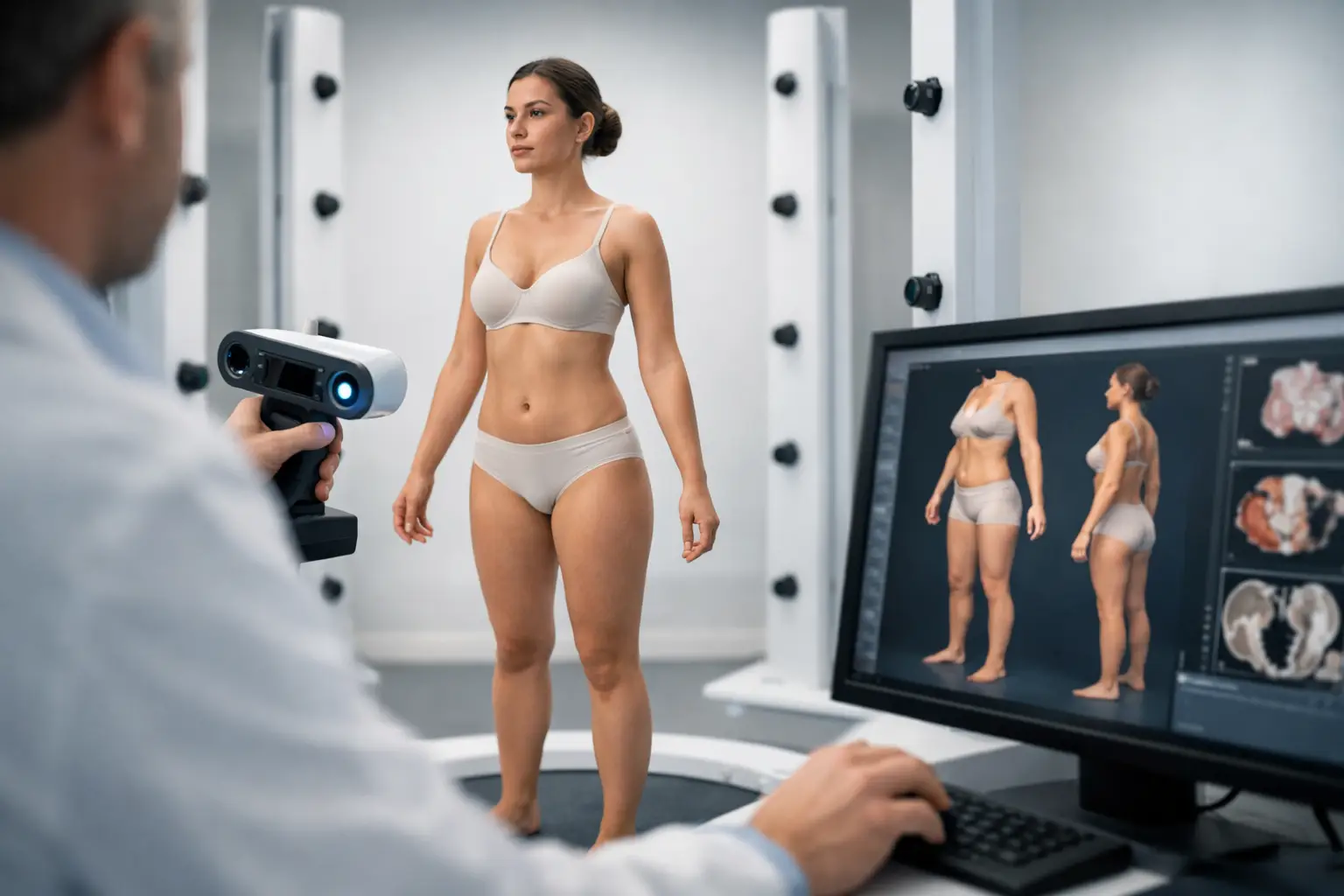
3D body scanning adds a more complete layer of truth. It captures the patient’s external anatomy as a digital model that can be measured, reviewed, and compared over time. That matters in aesthetic procedures, reconstructive work, orthotics, prosthetics, and any case where surface geometry influences decision-making.
The practical value is immediate. A surgeon can assess proportion with more context. A patient can see proposed changes in a way that feels more concrete. A clinic can standardize documentation instead of relying on inconsistent photo setups. The result is not magic. It is better spatial data.
That said, this is not a replacement for clinical judgment, CT, MRI, ultrasound, or other medical imaging where internal anatomy drives the case. 3D body scanning is strongest when external form is central to planning. Used in the right setting, it improves visibility. Used in the wrong setting, it can be mistaken for a full diagnostic system, and it is not that.
What 3D body scanning actually improves before surgery
The first improvement is communication. Many pre-op conversations break down because the patient and clinician are looking at the same issue from different mental models. The surgeon sees anatomical constraints. The patient sees a desired outcome. A 3D model creates a shared reference point. That reduces ambiguity.
The second improvement is baseline accuracy. A digital body scan can capture dimensions, contour transitions, and asymmetries more consistently than manual notes alone. In procedures where millimeters affect fit, balance, or visual harmony, that consistency has real value.
The third improvement is longitudinal comparison. When clinics scan the patient before intervention and again during recovery, changes become easier to evaluate. That can support expectation management, outcome review, and internal quality control.
There is also a business advantage. Practices that adopt spatial workflows often move faster from consultation to decision because the planning process feels more tangible. Better clarity can shorten the back-and-forth that delays treatment acceptance.
Where it fits best in surgery planning
Not every specialty benefits equally, and that distinction matters.
In aesthetic surgery, 3D body scanning is especially useful because contour, symmetry, and visible form are central to both planning and patient satisfaction. Breast procedures, body contouring, facial work, and post-weight-loss surgery all involve external geometry that patients want to understand before committing.
In reconstructive surgery, the technology can support surface assessment and documentation, especially when tracking defects, asymmetry, or post-operative progression. It can also help teams communicate planned changes more clearly across consultations.
In orthotics and prosthetics workflows, body scanning becomes even more operational. The scanned geometry can inform fit, customization, and iterative adjustments. Here, speed and repeatability matter just as much as visualization.
In general surgery or cases driven primarily by internal pathology, the value may be narrower. External body scans can still support documentation and patient engagement, but they are not the main planning engine. This is where disciplined adoption matters. The strongest clinics use the technology where it creates measurable value, not where it simply looks advanced.
3D body scanning for surgery planning and patient trust
Patients do not buy procedures. They buy confidence.
A 3D scan gives the consult room a stronger decision framework. Instead of abstract descriptions, patients can review their own anatomy in three dimensions. That often leads to better questions, more grounded discussions, and fewer false assumptions about what surgery can realistically achieve.
This has an important trade-off. Better visualization can improve trust, but only if the clinic presents the scan responsibly. If a patient interprets a digital model or simulation as a guaranteed result, the technology starts to work against the practice. The scan should support informed consent, not oversell certainty.
The best use case is straightforward: show anatomy clearly, explain constraints honestly, and use visual tools to align expectations with what the procedure can actually deliver.
What clinics should evaluate before adopting a scanning workflow
The first question is accuracy at the use-case level. A clinic does not need the same capture standard for every application. Aesthetic consultation, prosthetic fitting, and post-op tracking all have different tolerance thresholds. Teams should evaluate whether the scan quality is sufficient for the decision being made.
The second question is workflow friction. If a system produces excellent data but takes too long to capture, process, or review, staff will avoid it. Adoption usually succeeds when scanning is fast enough to fit inside normal consult operations.
The third question is accessibility. Dedicated hardware has its place, but it also creates bottlenecks around cost, training, and deployment. Smartphone-based capture is changing that equation. When high-quality scanning can happen on devices teams already use, implementation gets much easier to scale.
The fourth question is platform strategy. Point solutions solve one problem. Infrastructure solutions connect scanning to broader visualization, modeling, measurement, and workflow needs. That distinction matters more over time. The clinics investing seriously in spatial technology are not looking for isolated demos. They are building digital capability.
This is where companies like MagiScan have a strategic advantage. The market is moving beyond stand-alone scanning apps toward integrated spatial AI ecosystems that support capture, processing, and applied use across verticals, including medical workflows.
The smartphone shift is changing the category
For years, 3D capture in clinical environments carried a hidden assumption: serious scanning required specialized equipment, specialized operators, and specialized budgets. That assumption is breaking.
Smartphone-based scanning, powered by stronger computer vision and AI reconstruction, is expanding access to production-ready 3D capture. That does not mean every phone scan is instantly suitable for every medical application. It means the barrier to entry is collapsing fast, and the quality curve is rising.
That matters commercially. As capture becomes easier, more clinics can test workflows, train staff, and build repeatable use cases without large upfront infrastructure bets. It also matters strategically. Once spatial capture becomes mobile, the real differentiator shifts from hardware ownership to software intelligence, workflow design, and vertical execution.
In surgery planning, that opens the door to more frequent scans, better patient records, and broader use across locations and teams. Instead of treating 3D capture as a rare event, clinics can treat it as a routine layer of pre-op and post-op documentation.
The limits are real, and that is a good thing
Hype hurts adoption when it ignores constraints.
3D body scanning depends on good capture conditions. Lighting, patient positioning, movement, clothing, and operator technique all affect output quality. Standardization matters. A sloppy scan can create false confidence, which is worse than no scan at all.
There are also privacy and data governance issues. Body scans are sensitive patient data. Clinics need secure storage, controlled access, and clear consent practices. The more useful these models become, the more seriously practices need to treat them as part of the medical data environment.
And there is the interpretation gap. A scan is data, not judgment. It can improve planning, but it still needs an experienced clinician to decide what matters and what does not.
What comes next
The future of surgery planning is not just better imaging. It is editable reality.
As 3D body scanning merges with AI, simulation, and AR visualization, the planning process becomes more interactive and more scalable. Consultations become clearer. Documentation becomes richer. Teams make decisions with stronger spatial context. Over time, the winners will be the practices that treat 3D capture not as marketing theater, but as operational infrastructure.
That is the real opportunity in 3d body scanning for surgery planning. It gives clinicians a better way to see, explain, and prepare before treatment begins. When the technology is accurate enough, fast enough, and integrated into the right workflow, it stops feeling experimental. It starts feeling inevitable.
The smartest move now is not to ask whether spatial tools belong in pre-op planning. It is to ask where they create the most value first, then build from there.